DENTAL ANATOMY
Definition:
Dental Anatomy is the science that deals with the morphology of the structures and contours of the teeth, their relationship and immediately associate parts.
The parts of tooth are includes -
i. crown
ii. neck
iii. root
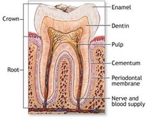
Figure: Normal Tooth anatomy
Morphological component of the teeth:
i. Enamel: Enamel is the most hardest tissue of the body. The thickness of enamel on tooth varies from 2.5mm to 0.5mm. The color of the enamel form yellowish white to grayish white. It form by ameloblost cell.
ii. Dentin : Dentin provide the bulk and general form of the tooth. It determine the shape of the crown. The dentin ranges and thickness from 3mm to 10mm.
iii. Cementum : It is the mineralized dental tissue covering the root of human teeth. Human cementum is avascular.
iv. Pulp : The pulp occupies the center of each teeth. It content nerve and blood vessel and specialized cell such as odontoblast, fibroblast and mesenchoymal cell.
Functions related to tooth:
The teeth are divided in groups corresponding to their functions. The eight incisors serve to incise or cut the food. The four cuspids serve to pierce and hold the food. The eight bicuspids serve to pierce the food and also to commnicate the foods. The twelve molars serve to grind the food and to communicate the food.
Dental Anatomy is the science that deals with the morphology of the structures and contours of the teeth, their relationship and immediately associate parts.
The parts of tooth are includes -
i. crown
ii. neck
iii. root
Figure: Normal Tooth anatomy
Morphological component of the teeth:
i. Enamel: Enamel is the most hardest tissue of the body. The thickness of enamel on tooth varies from 2.5mm to 0.5mm. The color of the enamel form yellowish white to grayish white. It form by ameloblost cell.
ii. Dentin : Dentin provide the bulk and general form of the tooth. It determine the shape of the crown. The dentin ranges and thickness from 3mm to 10mm.
iii. Cementum : It is the mineralized dental tissue covering the root of human teeth. Human cementum is avascular.
iv. Pulp : The pulp occupies the center of each teeth. It content nerve and blood vessel and specialized cell such as odontoblast, fibroblast and mesenchoymal cell.
Functions related to tooth:
The teeth are divided in groups corresponding to their functions. The eight incisors serve to incise or cut the food. The four cuspids serve to pierce and hold the food. The eight bicuspids serve to pierce the food and also to commnicate the foods. The twelve molars serve to grind the food and to communicate the food.
Type of Dentition
i. Primary dentition or Deciduous dentition
ii. Mixed dentition
iii. Permanent dentition
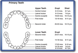
Figure:Primary teeth anatomy
Numbers of tooth:
Primary dentition ------- 20 in number. Eruption start at 6 month of age.
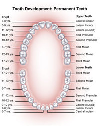
Permanent dentition-----32 in number. Eruption start at 6 years of age.
Figure: Permanent tooth anatomy
Eruption time of deciduous teeth
Lower central incisors – 6 to 7 months
Upper central incisors – 7 to 8 months
Lower lateral incisors – 7 to 8 months
Upper lateral incisors – 8 to 9 months
First deciduous molars – 15 to 16 months
Canine - 16 to 20 months
Second deciduous molar – 20 to 24 months
Eruption time of permanent teeth:
First molar – 6 yrs
Central incisors – 7 yrs
Lateral incisors - 8 yrs
First bicuspids – 10 yrs
Second bicuspids – 11 yrs
Third molars – 17 to 25 yrs
